Ultrasound technology has revolutionized the way we look inside the human body — safely, accurately, and painlessly. It plays a key role in diagnosing various health conditions, tracking pregnancy progress, and evaluating organ function. Among the latest advancements in ultrasound imaging are 3D and 4D ultrasound scans, which provide detailed and realistic visuals that go far beyond traditional 2D scans.
At Sonobeat Heart Care and Sonography Centre in Aurangabad, patients benefit from advanced imaging services, including 3D/4D sonography, Doppler studies, 2D Echo, and specialized cardiology and pregnancy scans — all performed with precision and care.
Let’s understand the difference between 3D and 4D ultrasound scans, their benefits, and when each is recommended.
What Is an Ultrasound Scan?
An ultrasound scan (also known as sonography) uses high-frequency sound waves to create real-time images of the inside of the body. Unlike X-rays or CT scans, ultrasounds don’t use radiation, making them completely safe — especially for pregnant women and infants.
The images are generated by a small handheld device called a transducer, which sends and receives sound waves. These waves bounce off internal tissues and organs, and the reflected signals are converted into live images on a monitor.
Ultrasound is widely used for:
- Pregnancy scans to monitor fetal growth and health
- Heart examinations like 2D Echo and Doppler tests
- Abdominal and pelvic imaging
- Detecting vascular diseases and organ abnormalities
Understanding 2D, 3D, and 4D Ultrasound Scans
2D Ultrasound – The Traditional Scan
2D ultrasound is the most common and basic type of scan. It produces flat, black-and-white, two-dimensional images that show a slice of what’s happening inside the body.
In pregnancy, 2D ultrasound helps:
- Confirm pregnancy
- Estimate the due date
- Monitor fetal heartbeat and movements
- Check for abnormalities
While 2D images are very informative, they may not always provide the depth or clarity needed for advanced evaluation — this is where 3D and 4D scans come in.
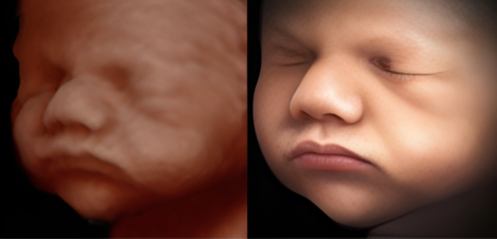
3D Ultrasound Scans – A New Dimension in Imaging
A 3D ultrasound creates three-dimensional still images by capturing multiple 2D images from different angles and combining them using advanced computer software.
In simple terms, it gives a lifelike, detailed image of your baby or the organ being examined.
Applications of 3D Ultrasound:
- Pregnancy:
3D ultrasound allows parents to see a more realistic image of their baby’s face, hands, and body. It helps detect certain congenital anomalies like cleft lip, spine defects, or limb abnormalities with greater clarity. - Gynecology:
Used to assess uterine abnormalities, ovarian cysts, or fibroids. - Cardiology:
Provides detailed views of heart structures, helping cardiologists plan accurate treatment.
Advantages of 3D Ultrasound:
- Produces clearer, more defined images
- Helps detect structural anomalies early
- Offers a better emotional connection for expecting parents
- Enhances diagnostic accuracy for doctors
4D Ultrasound Scans – Real-Time Motion in 3D
While 3D scans capture static images, 4D ultrasound adds the element of motion — the fourth dimension being time. It shows real-time video footage of what’s happening inside the womb or the body.
In pregnancy scans, this means you can actually watch your baby move, smile, yawn, or stretch in real time — an unforgettable bonding experience for parents.
Applications of 4D Ultrasound:
- Pregnancy:
Observe fetal movement and development dynamically. - Cardiology:
Evaluate heart motion and blood flow with precision. - Vascular studies:
View live blood circulation patterns using 4D Doppler imaging.
Advantages of 4D Ultrasound:
- Provides moving, live visuals for better analysis
- Enhances fetal assessment and diagnosis
- Offers a more interactive, emotional experience for parents
- Useful for detailed study of organ or heart motion
3D vs. 4D Ultrasound – Key Differences at a Glance
| Feature | 3D Ultrasound | 4D Ultrasound |
| Type of Image | Still, 3D image | Moving, real-time 3D image |
| Experience | Static view of structures | Dynamic video of movement |
| Use in Pregnancy | Detects structural defects | Observes live fetal activity |
| Diagnostic Value | High clarity | High clarity + motion analysis |
| Technology | Multiple 2D slices combined | Continuous 3D imaging over time |
Both 3D and 4D scans are safe and non-invasive, using the same sound wave principles as 2D scans. The main difference lies in the level of detail and real-time visualization.
When Are 3D and 4D Ultrasounds Recommended?
- Between 26–32 weeks of pregnancy:
This is the ideal time for 3D and 4D ultrasounds since the baby’s facial features are developed, and there’s enough amniotic fluid for clear imaging. - For cardiac and vascular evaluations:
When doctors need a dynamic view of the heart or blood flow, 4D Doppler imaging offers unmatched accuracy. - For structural analysis:
When a clearer understanding of abnormalities or tissue formation is needed.
Always consult your doctor before opting for a scan — they’ll recommend the right type based on your condition and stage of pregnancy.
Safety of 3D and 4D Ultrasound Scans
A common question among expectant parents is: Are 3D and 4D ultrasounds safe?
Yes — both are completely safe when performed by trained professionals.
At Sonobeat Heart Care and Sonography Centre, scans are conducted using state-of-the-art ultrasound machines under the supervision of experienced radiologists and cardiologists. The procedures are quick, comfortable, and safe for both mother and baby.
No radiation or harmful effects are involved — just sound waves that provide incredible insight into your health.
3D and 4D Ultrasounds at Sonobeat Heart Care and Sonography Centre, Aurangabad
At Sonobeat Heart Care and Sonography Centre, patients receive accurate, high-resolution imaging in a comfortable and patient-friendly environment.
The center specializes in:
- 3D/4D Pregnancy Scans: For detailed fetal evaluation and bonding moments.
- 2D Echo & Doppler Studies: For comprehensive heart and vascular health assessment.
- Fetal Echocardiography: Early detection of congenital heart issues.
- Cardiology Consultations: Expert care for heart-related conditions.
Every scan is performed with precision, ensuring both diagnostic clarity and patient comfort. The team is led by highly skilled professionals committed to quality care and compassion.
Why Choose Sonobeat Heart Care and Sonography Centre?
- Advanced 3D/4D ultrasound technology
- Expert team of cardiologists and sonographers
- Comfortable and hygienic setup
- Quick, reliable, and affordable diagnostic services
- Trusted by families across Aurangabad for pregnancy and cardiac care
Whether you’re an expectant parent eager to see your baby’s first smile or someone needing a detailed cardiac evaluation, Sonobeat Heart Care and Sonography Centre ensures a smooth, reassuring experience with world-class imaging accuracy.
Conclusion
Both 3D and 4D ultrasound scans mark a significant advancement in medical imaging. While 3D provides detailed still images, 4D adds movement — making diagnosis more precise and experiences more memorable.
At Sonobeat Heart Care and Sonography Centre technology meets compassion to deliver the best care for every patient. Whether it’s monitoring your baby’s growth or understanding your heart’s health, expert hands ensure every scan contributes to a healthier, happier future.